X-rays are a common part of most visits to the dentist or orthodontist. But did you know this technology wasn’t widely used for dental purposes until as recently as the 1950s?
Our team at glassortho.com can tell you how these x-rays work (because it’s cool) and, more importantly, answer the question, “Why do orthodontists use x-rays?”
What Did Orthodontists Do Before X-Rays?
Traditional methods and clinical examinations were used prior to the wide availability of X-ray technology in offices. Orthodontists could take exterior photographs to document and track a patient’s condition during treatment, but these couldn’t provide internal information. The closest they could get was through the use of something called panoramic radiographs, which are wide format, less detailed images than modern x-rays.
Other methods included bite analysis and taking molds and impressions of the mouth. They would also take plaster models at regular intervals to measure progress in the teeth and jaw during orthodontic treatment.
In other words, it wasn’t nearly as simple as pressing a button.
A Transparent Explanation
Dental x-rays are also called radiographs. They are a common diagnostic tool that uses electromagnetic radiation to take pictures of bones, teeth, and surrounding oral and facial structures. Curious how they work? Let’s take a look behind the scenes:
- The X-ray machine generates bursts of radiation. A dental assistant will usually be operating this device prior to Dr. Glass entering.
- You’ll be covered with something called a lead apron. It blocks your vital organs from ionizing radiation. Usually, it will just cover your chest and torso. It should also be noted that modern X-ray tech limits exposure to radiation much better than it used to.
- A digital sensor is placed inside the patient’s mouth and captures images. It can be positioned to either take photos of the whole mouth or focus on areas of concern. In the past, x-ray film was used for this purpose and would be developed in a lab.
- Once the machine is activated, a focused beam of exposure is directed toward the sensor. Softer tissues like gums and cheeks allow more x-rays to pass through, creating darker areas on the image, while teeth and bones absorb more x-rays due to their density, resulting in lighter areas on the final image.
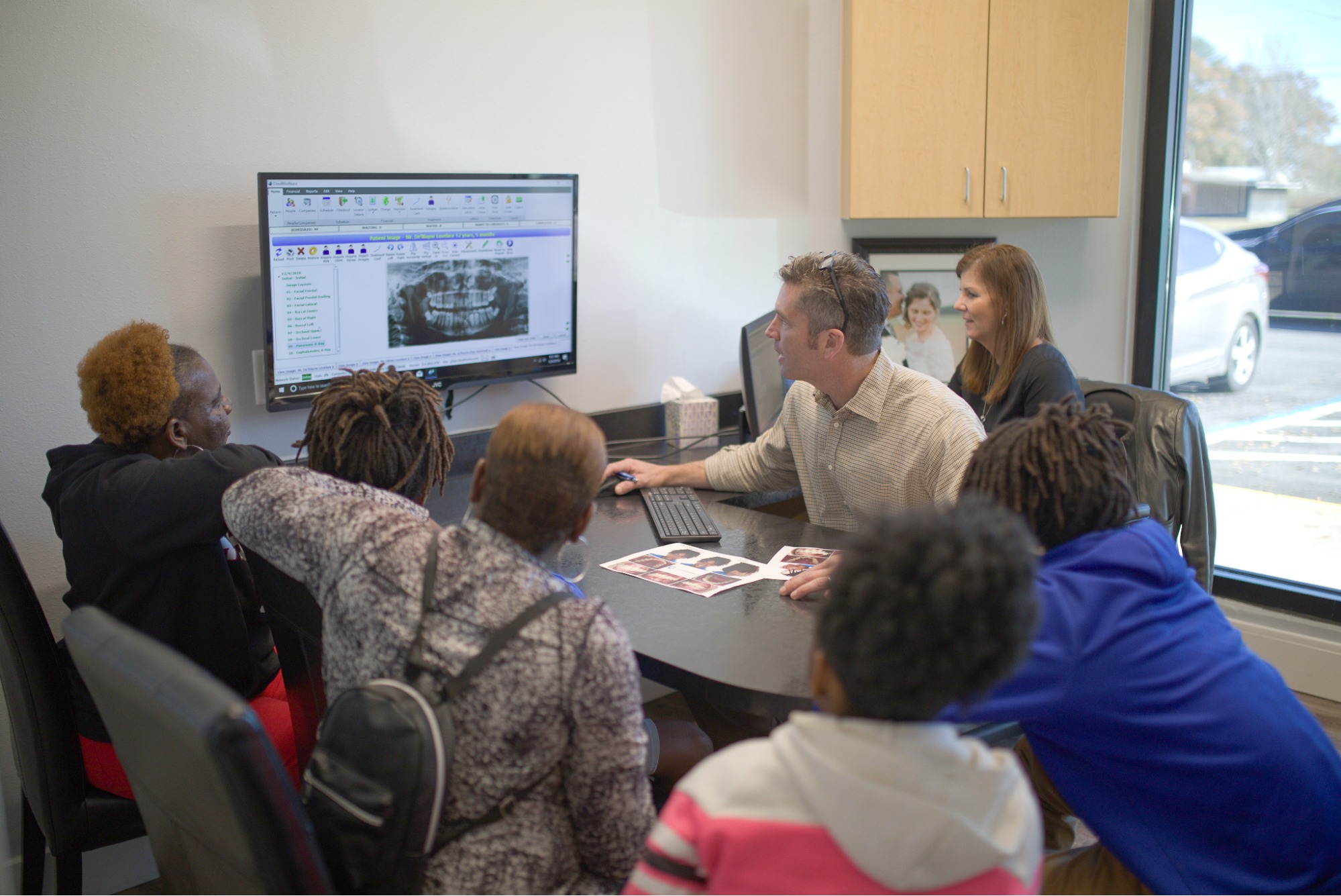
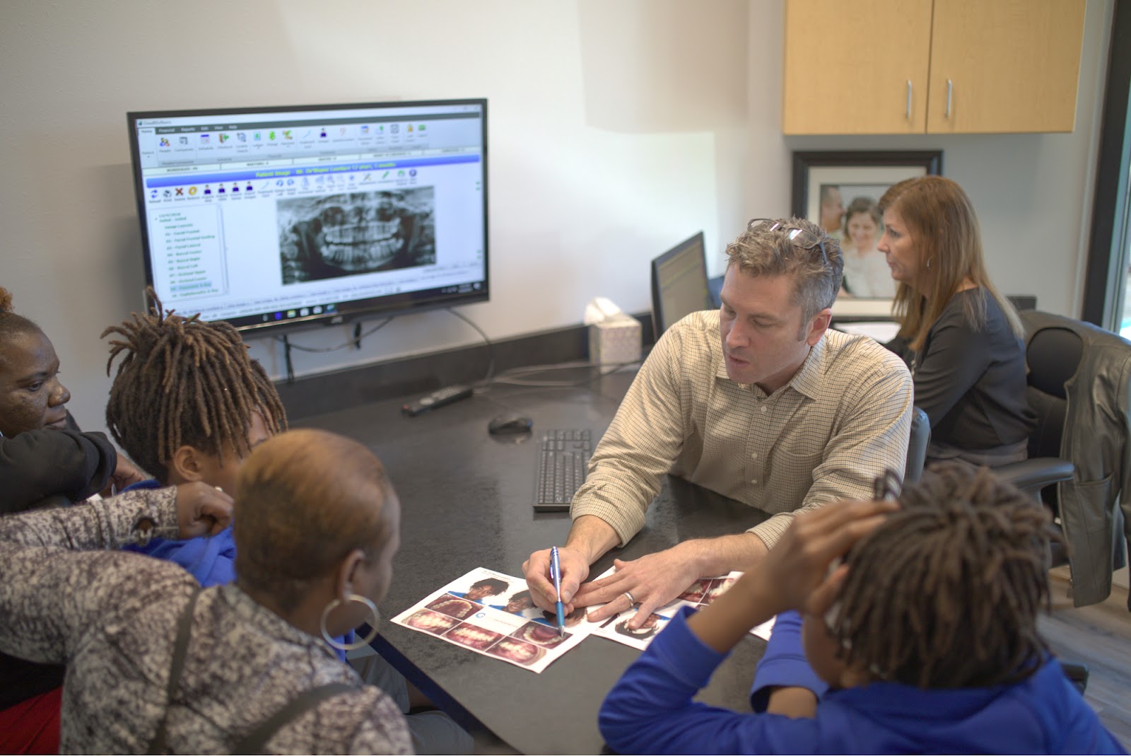
- The image appears on a screen and is then closely examined by Dr. Glass.
- Intraoral x-rays feature the inside of the mouth and make small areas visible.
- Extraoral x-rays feature the whole oral and facial structure, used for a broader view of the head and jaw when necessary.
Why We Use X-Rays
Now that you know how it all pans out, we can talk about why and how Dr. Glass and our awesome team use them.
- Early Diagnosis of Orthodontic Issues: We’re able to better pinpoint orthodontic problems in their early onset stages—hopefully before they become visibly apparent. We can then use an interceptive treatment and potentially prevent issues from getting more severe.
- Assessment of Tooth and Jaw Position: The most vital part of this technology is that it provides us with detailed images of the teeth, jawbone, and surrounding structures, which are not visible during a regular oral examination. These images allow us to address the position, alignment, and growth of teeth and jaws in our patients across Alabama.
- Evaluation of Dental Development: We can easily keep records showing the development of teeth, most notably in children and adolescents, sometimes spanning years. Even if you’re coming from another office, we can use your most recent x-rays in addition to our own for reference. This also helps our team observe how permanent teeth are coming in and determine if there are any issues.
- Assessment of Bite Problems: Overbites, underbites, crossbites, and open bites (otherwise known as malocclusion) are conditions we treat all the time. By visualizing the upper and lower jaws and the alignment of teeth, we can confidently decide on treatment options.
- Identification of Impacted Teeth: Impacted teeth don’t erupt properly or are blocked from eruption. Monitoring for impacted teeth is vital for planning treatments, especially in cases where oral surgery is required. You might hear this phrase used when you’re getting your wisdom teeth removed (but we hope not!).
- Bone Health: Density and health of the jawbone aren’t something you always consider, but we do! When our treatment plan involves the repositioning of teeth, bones must be capable of supporting these adjustments.
- Treatment Verification: After your orthodontic treatment is complete, we can take a look and confirm that your teeth and jaws have been successfully realigned according to Dr. Glass’s plans!
You’ll Excel With glassortho.com!
So, next time you come in, you’ll have a better idea of what actually happens when you have that funny weighted blanket on and a sensor in your mouth! Our team is happy to take advantage of this technology when we treat you. We’re committed to serving the communities of Bay Minette, Daphne, Atmore, and Brewton! Schedule your free consultation now, or contact any of our offices with questions.